Explore our infrastructure
At LBIC, we provide access to a range of pioneering technologies. Our dedicated staff scientists can offer support throughout the entire process chain; from project design, experimental set-up and preparation, to imaging, data storage, analysis and visualization. Additionally, we can also offer training, support, workshops and seminars.

| Microscopy
The Microscopy Facility offers a wide range of techniques for imaging of samples on a variety of resolution ranges, including the nanometer range obtained by electron microscopy and the micrometer range obtained by different types of light microscopy.
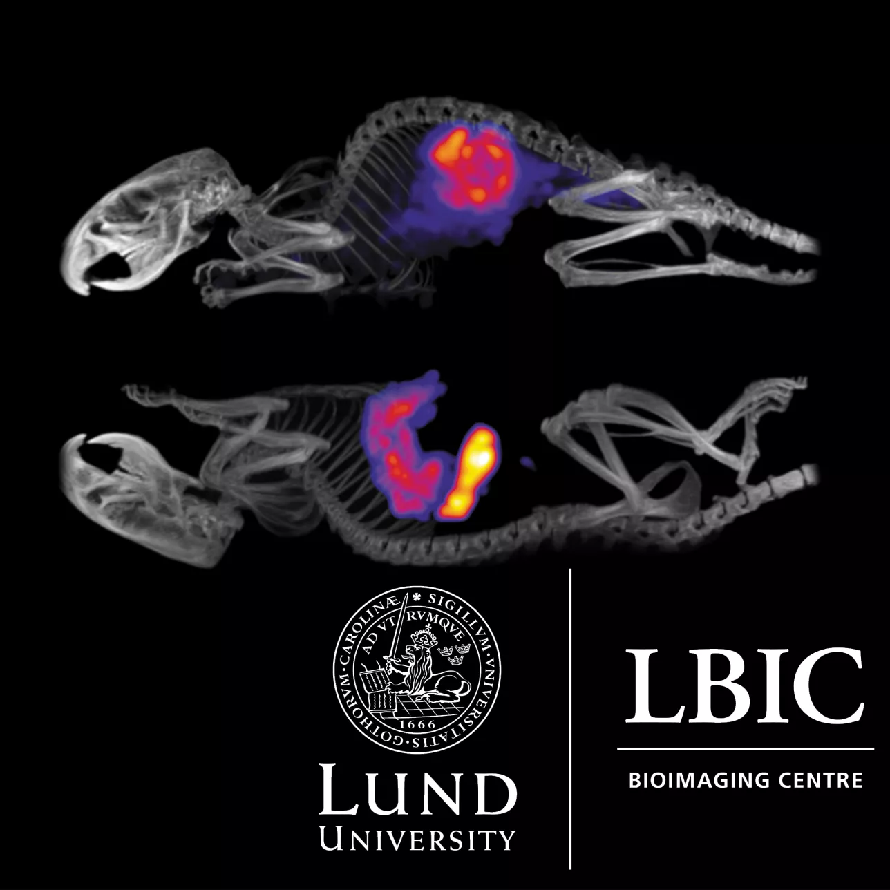
| Preclinical Nuclear Medicine
The Preclinical Nuclear Medicine Facility comprising of radiotracer techniques such as Positron Emission Tomography (PET) and Single-Photon Emission Computed Tomography (SPECT), which provide unique means for high-sensitivity studies of physiological function and molecular biodistribution. X-Ray Computed Tomography (CT), a technique to provide high-resolution anatomical imaging, often combined with PET and SPECT.
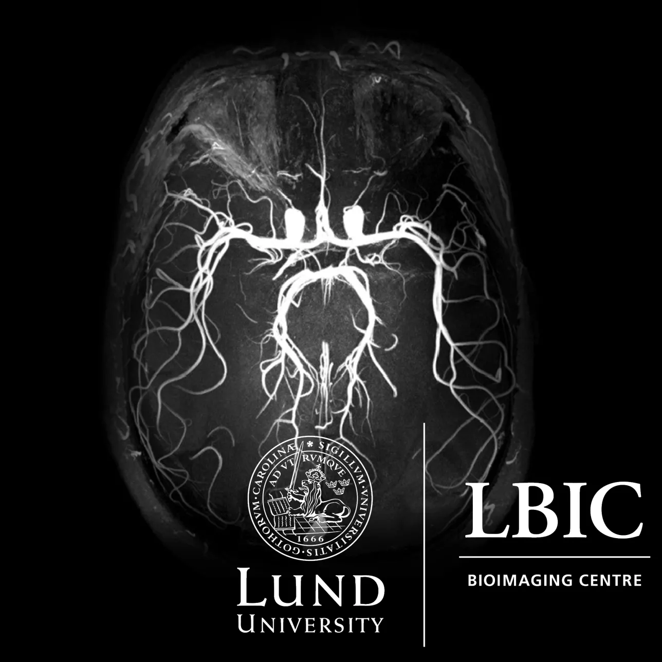
| Preclinical MRI
The Preclinical 9.4 Tesla Magnetic Resonance Imaging Facility offers a modern and robust imaging MRI system (9.4T) and staff with many years of practical experience in a wide variety of MR-applications. High-quality images of small animals and excised tissue can be obtained. The availability of a so-called cryo-coil further boosts the image quality for mouse imaging and small ex-vivo samples. Advanced MRI analysis of data is also available on request.
| Analysis & Visualization
The Analysis & Visualization Facility helps in the usage of image analysis software packages, image analyzation, production of 3D surface models, animations etcetera.
| Clinical MRI
The Clinical 1.5/3 Tesla Magnetic Resonance Imaging Facility comprises a variety of tools ranging from high-resolution anatomical structure visualization to functional studies and even molecular-level description of metabolic events in the cell.
| National 7 Tesla Facility
The National 7 Tesla Facility for ultra-high-field human magnetic resonance imaging research can offer equipment for brain, knee, wrist, breast and abdominal imaging, fMRI stimuli and eye tracking as well as clinical devices, such as contrast injector and ventilator. The facility is open for researchers from all Swedish universities.