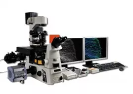
Advanced light microscopy
LBIC gives researchers access to 8 advanced light microscopes, all placed at Biomedicinskt Centrum (BMC).
Confocal laser scanning microscopes
Nikon A1RHD Confocal
Motorized confocal laser scanning microscope equipped with both a high-resolution galvano scanner and a high-speed (15 fps) 1024 x 1024 resonant scanner capable of capturing high-quality confocal images of tissues, cells and molecular events at high speed and enhanced sensitivity. This system is also equipped with a spectral detector.

Key features:
- Laser lines: 405 nm, 488 nm, 561 nm, and 640 nm
- Objectives: 4x, 10x, 20x, 40x & 60x oil
- Detectors: 2 GaAsP PMT (for 488 and 561 lines), 2 PMT (for 405 and 640 lines) and 1 GaAsP PMT for spectral detection (400-720nm detection range)
- Resonant scanner (15 fps) 1024 x 1024 pixel scan size
- Piezo Z-drive (NanoDrive, MadCity labs) for fast Z-stacks
- Software: NIS-Elements
Nikon A1+ Confocal
Motorized confocal laser scanning microscope equipped with a high-resolution galvano scanner capable of capturing high-quality confocal images of tissues, cells and molecular events at enhanced sensitivity.
Key features:
- Laser lines: 405 nm, 488 nm, 561 nm, and 640 nm
- Detectors: 2 GaAsP PMT (for 488 and 561 lines), 2 PMT (for 405 and 640 lines)
- Objectives: 10x & 20x
- Software: NIS-Elements
To find out more about the confocal technique go to:
http://www.microscopyu.com/articles/confocal/confocalintrobasics.html (Opens in new window)
Wide-field fluorescence microscopes
Nikon Ti2 FM
Motorised wide-field fluorescence microscope with large field of view equipped with a monochrome sCMOS camera as well as a color camera. The microscope is owned by the Iben Lundgaard group and available to LBIC users 20h per week. Wide-field fluorescence microscopy is an extensively used microscopy technique that illuminates the entire objective field of view (FOV) with excitation light and collects the resulting fluorescence signal. Best suited for thin stained or fluorescently labeled thin tissue sections or cell cultures.

Key features:
- LED illumination lines: 405nm, 488nm, 561nm, and 647nm.
- Cameras:
- Monochrome 16.25 MpNikon DS-Qi2 sCMOS, active pixels (w*h) 2424x2424. Maximum quantum efficiency 77%.
- Color 5Mp Sony Pregius CMOS, active pixels (w*h) 2448 x 2048.
- Objectives: 4x, 10x, 20x & 40x
- Software: NIS-Elements
Nikon Ti2 BSL2 live-cell FM
Motorised wide-field fluorescence microscope with large field of view in a cage incubator equipped with a monochrome sCMOS camera. The microscope is placed in a Bio safety level 2 (BSL2) lab enabling study of Class 2 pathogens and host-pathogen interaction studies. The system owned by the Pontus Nordenfelt group and available to LBIC users 8h per week.

Key features:
- LED illumination lines: 405nm, 488nm, 561nm, and 647nm.
- Camera: Monochrome 16.25 Mp Nikon DS-Qi2 sCMOS, active pixels (w*h) 2424x2424. Maximum quantum efficiency 77%.
- Objectives: 10x, 20x, 40x and 100x oil
- Placed in Bio Safety Level 2 lab
- Cage incubator for stable long term live-cell imaging
- Software: NIS-Elements with JOBS scripting
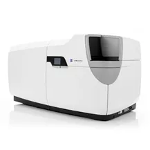
Zeiss CD7 automated live cell Microscope
ZEISS Celldiscoverer 7 is an automated microscopy platform designed for live cell imaging and analysis. It combines a range of advanced imaging technologies enabling high-resolution imaging of live cells and tissues over extended periods of time. Its features include LED-based illumination, sensitive camera, and a range of imaging modes, including fluorescence, brightfield, and phase contrast. The system also has integrated environmental control, allowing for stable and consistent imaging conditions and is coupled with an automated incubator and plate handler. The CD7 is placed at BMC floor A12 and is shared with the Filipe Pereira group and available to LBIC users 20h per week.
Key features:
- LED illumination lines: 385nm, 420nm, 470nm, 520nm, 567nm, 590nm and 625nm
- Camera: monochrome 6Mp Axiocam 506, active pixels (w*h) 2752 x 2208.
- Objectives: 5x, two 20x and 50x water immersion. System has a magnification changer (0.5x, 1x and 2x) which means that each objective can be used for three different magnifications
- Integrated environmental control for stable long term live-cell imaging
- Thermo fisher scientific Cytomat 2C automated incubator
- PAA S-LAB plate handler
To find out more about fluorescence microscopy go to:
https://www.microscopyu.com/techniques/fluorescence/introduction-to-fluorescence-microscopy (Opens in new window)
Total internal reflection fluorescence microscopy
Nikon TiE TIRF
Motorized Nikon TiE TIRF microscope with stage-top temperature incubator for short term live-cell TIRF imaging. Total internal reflection microscopy (TIRF microscopy) uses the phenomenon of total internal reflection to create an evanescent wave that only illuminates a thin area near the interface between the glass coverslip and the aqueous sample. This gives superior signal to noise images compared to conventional wide-field fluorescence but is only suitable for studying events taking place at or very near (100-200nm) the coverslip. The system is equipped with an Photometrix Prime95B sCMOS camera (owned by the Vinay Swaminathan group), which enables imaging at very high sensitivity and speed. The high speeds of the cameras allow for tracking of low-light dynamic events within a live cell in the “TIRF zone”.
Key features:
- Laser lines: 405nm, 488nm Argon with 457nm, 477 and 515nm side lines, 561nm, and 647nm.
- Camera: Photometrix Prime95B sCMOS
- Objectives: 10x, 20x and 100x TIRF
- Incubator: Stage top incubator with temperature control.
- Software: NIS-Elements
To find out more about the TIRF technique:
http://www.microscopyu.com/articles/fluorescence/tirf/tirfintro.html (Opens in new window)
Light Sheet Microscopes
Light sheet microscopy (LSM) differs from conventional light microscopy by having the illumination and detection optical pathways decoupled. In LSM, the sample is illuminated by a thin sheet of light (low- or sub-micrometer range) and the emitted light is detected orthogonally to the plane of illumination. Because of this, LSM imaging significantly reduces out-of-focus emitted light and an image of the illuminated plane can be quickly recorded using a CCD or CMOS camera. Optical sectioning and generation of 3D-images is there for possible using LSM by moving the sample through the light sheet or vice versa.
Furthermore, as only the optical plane of interest is illuminated in LSM, this technique offers reduced photo-bleaching and -toxicity compared to traditional light microscopy, where the whole detection path is illuminated. Due to the rapid imaging, high lateral and axial resolution, and reduced sample illumination associated with LSM, this technique is a great choice for imaging large biological samples (especially in combination with optical clearing techniques) and/or live imaging.
LBIC staff can help with optical clearing. Contact us to discuss alternatives.
To find out more about light sheet microscopy go to:
https://www.microscopyu.com/techniques/light-sheet/light-sheet-fluorescence-microscopy (Opens in new window)
Ultramicroscope Blaze light sheet
Key features:
- Light sheet type: Gaussian beam
- Lasers and emission filters:
- 405nm - 460/40
- 488nm - 525/50
- 561nm - 595/40
- 639nm - 680/30
- 785nm - 810/90
- Objectives:
- 1.1x/0.1, with 2 dipping caps ranging from RI (1.33-1.56)
- 4X/0.35 with 3 dipping caps (Water, Clarity, Eci)
- 12x/0.53 3 dipping caps (Water, Clarity, Eci)
- Camera: PCO 4.2 MP sCMOS camera, 2048×2048 pixels (w×h). Maximum quantum efficiency 82%.
- Sample cuvettes:
- Standard Cuvette – for single or multiple tissue samples (organoids to eg mice/rat organs)
- XL cuvette - for large samples (up to small mice)
MSquared Aurora Light sheet
Key features:
- Light sheet type: Airy beam
- Lasers and emission filters:
- 405nm - 450/50
- 457nm - 480/30
- 488nm - 520/40 & 535/30
- 561nm - 605/52
- 594nm - 624/40 & 632/60
- 647nm - 697/60
- 730nm - 750LP
- Camera: Hamamatsu Orca Flash4, 4.2 MP sCMOS camera, 2048×2048 pixels (w×h). Maximum quantum efficiency 82%.
- Objectives: Navitar Immersion objectives (~16x magnification) compatible with aqueous clearing solutions (e.g. CUBIC, Clarity, and ScaleS) and hydrophobic solutions (e.g. ethyl cinnamate)
- Sample chambers suitable for sample sizes from organoids to rat organs.